Adult T-cell leukemia/lymphoma
Alerts and Notices
Important News & Links
Synopsis

Adult T-cell leukemia / lymphoma (ATLL) is an aggressive peripheral T-cell neoplasm associated with human T-cell lymphotropic virus type 1 (HTLV-1) infection. HTLV-1 is endemic in southwestern Japan, the Caribbean, and parts of central Africa. It is acquired through blood transfusions, sexual intercourse, or breastfeeding. ATLL occurs exclusively in adults (average patient age is 58 years old) with a male to female ratio of 1.5:1.
Five clinical variants exist on a spectrum: acute (most common), lymphomatous, chronic, extranodal primary cutaneous variant, and smoldering. They vary based on their presentation and prognosis.
The acute variant (~50% of cases) is characterized by a high WBC count with lymphocytosis, generalized lymphadenopathy, hepatosplenomegaly, high lactate dehydrogenase (LDH), eosinophilia, cutaneous findings, and hypercalcemia (with or without lytic bone lesions). Bone marrow involvement is seen in up to one-third of cases. It has a poor prognosis, and the median survival is approximately 8 months.
The lymphomatous variant (~20% of cases) presents with significant lymphadenopathy and absence of peripheral blood involvement. The median survival is 11 months.
The chronic variant (~10% of cases) usually presents with an exfoliative dermatitis, mild lymphadenopathy, and lymphocytosis without internal organ involvement and has a median survival of 32 months, while the smoldering variant (~10% of cases) shows a normal WBC count with presence of circulating neoplastic cells and a median survival of 55 months. Occasionally, skin involvement is the only finding in the smoldering subtype. The primary cutaneous variant is rare and presents primarily with cutaneous involvement.
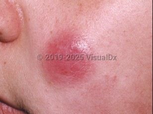
Cutaneous manifestations can be present in any of the variants, as well as pulmonary lesions. Cutaneous findings are seen in around 40%-70% of patients with ATLL. These are heterogeneous and may be specific or nonspecific. Specific presentations include a widespread papular eruption, patches and plaques (may resemble mycosis fungoides), the nodular-tumoral variant, erythroderma, and, rarely, a purpuric variant.
Patients with ATLL are immunosuppressed and at risk of developing opportunistic infections, thus giving rise to the nonspecific cutaneous features such as the presence of herpes zoster, molluscum contagiosum, dermatophytosis, or warts. There is similarly an increased risk of systemic opportunistic infections.
Codes
ICD10CM:
C91.00 – Acute lymphoblastic leukemia not having achieved remission
SNOMEDCT:
110007008 – Adult T-cell leukemia/lymphoma
C91.00 – Acute lymphoblastic leukemia not having achieved remission
SNOMEDCT:
110007008 – Adult T-cell leukemia/lymphoma
Look For
Subscription Required
Diagnostic Pearls
Subscription Required
Differential Diagnosis & Pitfalls

To perform a comparison, select diagnoses from the classic differential
Subscription Required
Best Tests
Subscription Required
Management Pearls
Subscription Required
Therapy
Subscription Required
References
Subscription Required
Last Reviewed:05/10/2023
Last Updated:05/11/2023
Last Updated:05/11/2023
Adult T-cell leukemia/lymphoma

