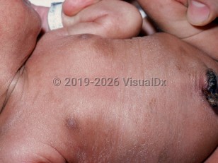
Fanconi anemia in Child
Alerts and Notices
Important News & Links
Synopsis

Fanconi anemia (FA) is a rare genetic syndrome that leads to loss of all formed elements of blood. Defects in several genes responsible for a DNA damage-activated signaling pathway give rise to FA in both autosomal and X-linked inheritance patterns. Individual hematologic cells are thus hypersensitive to DNA cross-linking agents, which leads to an increased rate of chromosome aberrations and cell death. This progressive degenerative disease results in a state of pancytopenia. Common associated abnormalities include congenital malformations of the kidney, heart, eye, central nervous system, and limbs. However, many are born without these congenital anomalies, which may delay diagnosis until childhood. Skin pigmentary changes may be the initial clue to the diagnosis.
Approximately 80% of individuals with the condition will have cutaneous manifestations. These include generalized hyperpigmentation, café-au-lait spots, and depigmented macules. Patients may also have hyperpigmentation secondary to iron overload from repeated blood transfusions. Skeletal abnormalities are very common, afflicting approximately 66% of affected patients. The most frequent of these are short stature, radial ray abnormalities, and scoliosis.
In addition, these patients are at increased risk of malignancy, most commonly nonlymphatic leukemia, with the increased risk of acute myeloid leukemia being as high as 15 000-fold. Other malignancies are observed with increased frequency, including head and neck squamous cell carcinomas, liver and brain tumors, esophageal carcinoma, and neuroepithelial tumors.
Patients typically die from infection, neoplasia, or hemorrhage. Immunodeficiency is not a prominent feature.
Note: Certain mutations in BRCA2 (also known as FANCD1) can cause a rare form of Fanconi anemia (subtype FA-D1) if inherited from both parents, and certain mutations in BRCA1 (also known as FANCS) can cause a different Fanconi anemia subtype if inherited from both parents.
Codes
ICD10CM:
D61.09 – Other constitutional aplastic anemia
SNOMEDCT:
30575002 – Fanconi's anemia
D61.09 – Other constitutional aplastic anemia
SNOMEDCT:
30575002 – Fanconi's anemia
Look For
Subscription Required
Diagnostic Pearls
Subscription Required
Differential Diagnosis & Pitfalls

To perform a comparison, select diagnoses from the classic differential
Subscription Required
Best Tests
Subscription Required
Management Pearls
Subscription Required
Therapy
Subscription Required
References
Subscription Required
Last Updated:01/16/2022
Fanconi anemia in Child

