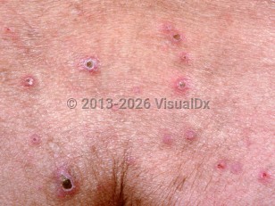
Reactive perforating collagenosis in Child
Alerts and Notices
Important News & Links
Synopsis

There is also a familial form of RPC, which has an onset in childhood. Familial RPC can persist throughout childhood and into adulthood, and generally presents on the hands and upper extremities. Lesions are induced by trauma.
The pathogenesis is unknown, but the familial disease is generally thought to be inherited in an autosomal recessive manner (although an autosomal dominant pattern has been observed in some families). Males and females are equally affected, and lesions generally start within the first few years of life in the inherited form.
The primary lesion is a skin-colored papule 1-3 mm in diameter that reaches a maximum size of about 6 mm within a month; 6-8 weeks later, it regresses spontaneously (often leaving temporary areas of hypopigmentation behind or slight scarring). There is a central keratotic plug seen in more advanced lesions that is firmly adherent and, if removed, results in bleeding. Koebner phenomenon may occur, resulting in new lesions that are often linearly arranged. Pruritus is often severe. There are often periods of disease remission and exacerbation.
Codes
L87.1 – Reactive perforating collagenosis
SNOMEDCT:
64036004 – Reactive perforating collagenosis
Look For
Subscription Required
Diagnostic Pearls
Subscription Required
Differential Diagnosis & Pitfalls

Subscription Required
Best Tests
Subscription Required
Management Pearls
Subscription Required
Therapy
Subscription Required
Drug Reaction Data
Subscription Required
References
Subscription Required
Last Updated:01/23/2022

