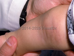
Tufted angioma
Alerts and Notices
Important News & Links
Synopsis

Tufted angioma, previously known as angioblastoma of Nakagawa, is a rare vascular tumor. The name describes the histologic appearance of "tufted" capillary infiltration of the dermis admixed with dilated lymphatic vessels. It is theorized to have a lymphatic origin.
Tufted angioma typically presents at birth (15% of cases) or within the first 5 years of life (50% of cases) as a violaceous plaque or nodule that may be painful. Rarely, it presents later in childhood or in early adulthood. There may be a slight male predominance.
After an initial period of growth, the tumor stabilizes and usually persists; in some patients, it may spontaneously, partially, or completely regress.
Three clinical patterns have been described: tufted angioma without complications, tufted angioma complicated with chronic coagulopathy without thrombocytopenia, and tufted angioma associated with Kasabach-Merritt phenomenon (KMP). KMP is a potentially life-threatening condition characterized by an enlarging vascular lesion, profound thrombocytopenia, consumptive coagulopathy, and often a microangiopathic hemolytic anemia.
Codes
ICD10CM:
D18.01 – Hemangioma of skin and subcutaneous tissue
SNOMEDCT:
254786000 – Tufted angioma of skin
D18.01 – Hemangioma of skin and subcutaneous tissue
SNOMEDCT:
254786000 – Tufted angioma of skin
Look For
Subscription Required
Diagnostic Pearls
Subscription Required
Differential Diagnosis & Pitfalls

To perform a comparison, select diagnoses from the classic differential
Subscription Required
Best Tests
Subscription Required
Management Pearls
Subscription Required
Therapy
Subscription Required
References
Subscription Required
Last Reviewed:08/11/2021
Last Updated:01/25/2022
Last Updated:01/25/2022
Tufted angioma

