Drug-induced nail pigment - Nail and Distal Digit
Alerts and Notices
Important News & Links
Synopsis

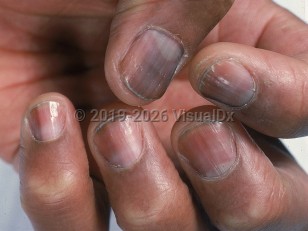
Cancer chemotherapeutic agents may result in melanonychia and leukonychia 1-2 months from the start of treatment. Doxorubicin can produce leukonychia and melanonychia within the same nail. Melanonychia is most often due to cyclophosphamide, doxorubicin, and hydroxyurea. Longitudinal bands, diffuse nail pigmentation, and associated skin pigmentation may be seen. The nail changes are due to activation of melanocytes within the nail matrix.
True leukonychia (due to faulty keratinization of the distal nail matrix) may be seen with treatment with antimetabolites including doxorubicin, cyclophosphamide, and vincristine. Apparent leukonychia (due to defects in nail bed blood flow) may also occur with chemotherapy. Some drugs, such as doxorubicin, can cause the occurrence of both leukonychia and melanonychia in the same nail.
Minocycline may occasionally cause abnormal pigmentation of the nails, and may also involve the skin, skin, teeth, mucosa, and sclera. It is thought to occur at cumulative doses greater than 100 g. It typically causes blue-black or slate-gray pigmentation of the proximal nail bed. Psoralen plus UVA (PUVA) therapy may result in longitudinal, transverse, or diffuse melanonychia, more often with 8-methoxypsoralen (8-MOP) than with 5-MOP.
Azidothymidine (AZT) may cause a number of different patterns of nail pigmentation including bluish-to-brown, diffuse nail discoloration, transverse or longitudinal banding, and a faint blue pigmentation of the lunulae.
Greenish florescence of nails has been reported after favipiravir, an antiviral medication given for COVID-19 infection.
Related topics: Longitudinal melanonychia, Leukonychia, Minocycline drug-induced pigmentation
Codes
L60.8 – Other nail disorders
T50.905A – Adverse effect of unspecified drugs, medicaments and biological substances, initial encounter
SNOMEDCT:
402632008 – Nail color change due to ingested drug
403302004 – Drug-induced leukonychia
Look For
Subscription Required
Diagnostic Pearls
Subscription Required
Differential Diagnosis & Pitfalls

Subscription Required
Best Tests
Subscription Required
Management Pearls
Subscription Required
Therapy
Subscription Required
Drug Reaction Data
Subscription Required
References
Subscription Required
Last Updated:11/22/2021

