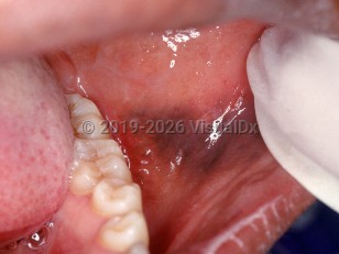
Multifocal or diffuse mucosal pigmentation - Oral Mucosal Lesion
Alerts and Notices
Important News & Links
Synopsis

Mucosal pigmentation caused by exogenous substances can be divided into two categories depending on the pigment: heavy metal pigmentation of systemic origin and medication-induced pigmentation. Heavy metal pigmentation may be caused by lead, arsenic, bismuth, or lead exposure from contaminated opium and often presents as a linear discoloration along the gingival margin (the "lead line" [Burton's line]). Even the bismuth present in Pepto-Bismol can cause a hyperpigmentation, although this is generally localized contact pigmentation on the dorsum of the tongue. It is believed that heavy metal ions excreted through the gingival crevicular fluid react with sulfites produced by gingival and periodontal bacteria, resulting in the deposition of heavy metal sulfides that are often black.
The medications that can cause drug-induced oral pigmentation include minocycline, antimalarials, clofazimine, and oral contraceptives. The drug or drug metabolites are pigmented substances that can be identified lying free or chelated to iron or melanin within the hard and/or soft tissues. Other drugs that supposedly cause oral pigmentation are likely related to a postinflammatory hypermelanosis from a lichenoid interface drug eruption or from direct damage to the mucosa (such as chemotherapeutic agents). Although tetracycline can cause intrinsic staining of the teeth and bone, it does not generally cause mucosal pigmentation.
Mucosal pigmentation caused by endogenous pigment is usually melanotic or hemosiderotic in origin, the latter from the breakdown of blood products. Melanotic pigmentation is seen in Peutz-Jeghers syndrome, McCune-Albright syndrome, hypoadrenal states (such as Addison disease), neurofibromatosis, LEOPARD syndrome, Laugier-Hunziker syndrome, and Carney complex, to name a few.
Codes
K13.79 – Other lesions of oral mucosa
SNOMEDCT:
249405005 – Oral pigmentation
Look For
Subscription Required
Diagnostic Pearls
Subscription Required
Differential Diagnosis & Pitfalls

Subscription Required
Best Tests
Subscription Required
Management Pearls
Subscription Required
Therapy
Subscription Required
Drug Reaction Data
Subscription Required
References
Subscription Required
Last Updated:05/02/2019

