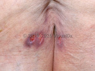
Typical sites of formation of a stage 3 pressure injury are the sacrum followed by the heels. A primary cause of pressure injury formation is immobility. Constant pressure for a time period of 2 hours is all that is required to initiate an ischemic event and to cause ulceration.
Other risk factors that predispose to ulcer formation include incontinence, nutritional deficits, old age, altered mental status, and malnutrition.
When examining the ulcer, observe the following specific points:
- Location on the body
- Stage of the ulcer
- Size of the ulcer, including depth, width, and length in centimeters.
- Wound bed – Appearance of the wound bed and the type of tissue visible. Observe the tissue color and whether it appears moist. The wound bed color of healthy granulating tissue is beefy-red and cobblestone-like. A red and smooth wound bed is indicative of clean but nongranulating tissue.
- Wound edges – Look carefully at the edge of the ulcer for evidence of induration, maceration, rolling edges, and redness.
- Skin around the edges of the ulcer – The periwound skin should be assessed for color, texture, temperature, and integrity of the surrounding skin.
- Drainage; exudate – If present, the color, amount, and presence of any odor.
- Presence of undermining, tunneling, or sinus tracts. Tunneling is a passage that extends from the wound through to subcutaneous tissue or muscle. Undermining is the destruction of tissue around the edge of that wound.
- Presence of necrotic tissue
- Presence or absence of pain
- Odor, if present or absent
- Medical device-related pressure injury (describes an etiology) – Results from the use of devices designed and applied for therapeutic purposes. Injury generally conforms to the pattern or shape of the device. Stage using the staging system.
- Mucosal membrane pressure injury – Found on mucous membranes with a history of a medical device in use at the location of the injury. Cannot be staged due to anatomy of tissue.


