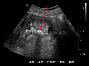
Renal calculus
Alerts and Notices
Important News & Links
Synopsis
As many as 80% of the stone disease in the United States are calcium oxalate or calcium phosphate stones. Calcium stones form when the concentration of these solutes (calcium, oxalate, or phosphate) exceed the solubility threshold of the urine in which they are dissolved. The solubility of these solutes is influenced by urine pH, the presence of other free ions, urinary organic solutes, and the presence of other crystals which provide nucleation sites for new crystal formation. When the urine is supersaturated and other factors are favorable, crystals form and precipitate, forming either stones inside of the renal pelvis or in submucosal plaques eponymously known as Randall plaques.
The primary risk factors for the development of renal calculi include male sex, increasing age (with a peak age of onset 20-40 years), obesity, European descent, high intake of animal protein, sodium, and fructose, a family history of stones, and living in the Southeast United States. Other, less common, risk factors include rare genetic conditions, gastric bypass surgery, and vitamin B6 (pyridoxine) deficiency.
Although asymptomatic stones are frequently discovered with radiographic imaging, the most common presentation is sudden-onset flank pain radiating to the groin, accompanied by nausea and vomiting. This colicky pain (renal colic) typically waxes over the course of 15-30 minutes and becomes steady, unrelenting, and unbearable. Patients may experience worsening paroxysms of pain lasting 20-60 minutes as the stone courses downward through the ureter and as the ureter spasms. If the stone's descent is arrested at the ureterovesical junction, patients may experience urinary frequency, dysuria, and urgency and are predisposed to the development of urinary tract infections both from the stone forming as a nidus for bacterial growth and from the mechanical urothelial trauma caused by the stone's movement. Most individuals with nephrolithiasis will also develop hematuria, particularly when passing a stone.
Pain from nephrolithiasis is thought to primarily be the result of renal capsular distention and varies depending on the location of the stone and the degree of obstruction caused by the stone. Stones that occlude the upper ureter or ureteropelvic junction invariably cause significant flank pain that is accompanied by severe costovertebral angle tenderness to palpation. As the innervation of the testicle is shared with the kidney, patients often describe radiation to the testicles or labia. When stones pass into the bladder, patients usually experience swift resolution of their pain.
Codes
N20.0 – Calculus of kidney
SNOMEDCT:
95570007 – Kidney stone
Look For
Subscription Required
Diagnostic Pearls
Subscription Required
Differential Diagnosis & Pitfalls

Subscription Required
Best Tests
Subscription Required
Management Pearls
Subscription Required
Therapy
Subscription Required
Drug Reaction Data
Subscription Required
References
Subscription Required
Last Updated:02/13/2022
 Patient Information for Renal calculus
 - Improve treatment compliance
- Reduce after-hours questions
- Increase patient engagement and satisfaction
- Written in clear, easy-to-understand language. No confusing jargon.
- Available in English and Spanish
- Print out or email directly to your patient


