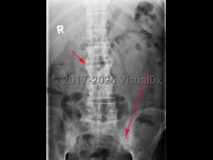
Ankylosing spondylitis
Alerts and Notices
Important News & Links
Synopsis
Ankylosing spondylitis is an autoimmune inflammatory disorder characterized by inflammation of the axial skeleton and peripheral joints. It typically begins in the second or third decade of life with a male-to-female prevalence of 2-3:1. There is a high concordance of ankylosing spondylitis in patients with haplotype human leukocyte antigen B27 (HLA-B27).
Patients most typically present with dull pain in the lower lumbar or gluteal region accompanied by early morning stiffness that improves with activity but not with rest. As the disease progresses, pain becomes persistent and bilateral and may worsen at night. Cervical ("chalk-stick") fractures may occur, especially after trauma. In addition to back pain, enthesitis (tenderness at the tendon or ligamentous insertion site to bone) may be common at the costosternal junction, spinous processes, iliac crests, greater trochanters, ischial tuberosities, tibial tuberosities, and heels. Approximately 30% of patients experience arthritis of peripheral joints other than the hips and shoulders. The most common extraarticular manifestation is acute anterior uveitis in up to 40% of patients. A large percentage of others may have inflammation of the terminal ileum or colon, although the majority of cases are asymptomatic. Only a minority of these patients progress to develop inflammatory bowel disease.
Physical examination findings reveal limited spinal mobility with limitation of anterior and lateral flexion / extension of the lumbar spine. Patients may also have limited chest wall expansion and tenderness at the tendinous insertion sites.
Codes
ICD10CM:
M45.9 – Ankylosing spondylitis of unspecified sites in spine
SNOMEDCT:
9631008 – Ankylosing Spondylitis
M45.9 – Ankylosing spondylitis of unspecified sites in spine
SNOMEDCT:
9631008 – Ankylosing Spondylitis
Look For
Subscription Required
Diagnostic Pearls
Subscription Required
Differential Diagnosis & Pitfalls

To perform a comparison, select diagnoses from the classic differential
Subscription Required
Best Tests
Subscription Required
Management Pearls
Subscription Required
Therapy
Subscription Required
References
Subscription Required
Last Updated:07/05/2022
Ankylosing spondylitis

