Contents

SynopsisCodesLook ForDiagnostic PearlsDifferential Diagnosis & PitfallsBest TestsManagement PearlsTherapyReferences

Information for Patients

View all Images (73)

Dermatofibroma in Adult
Other Resources UpToDate PubMed

Dermatofibroma in Adult

Contributors: Haya Raef MD, Belinda Tan MD, PhD, Sarah Hocker DO, Susan Burgin MD
Other Resources UpToDate PubMed

Synopsis

Dermatofibromas (DFs), or fibrous histiocytomas, are common, benign skin neoplasms composed of collagen, macrophages (histiocytes), capillaries, and fibroblasts. The etiopathogenesis is unknown, though some may arise at sites of trauma or insect bites. They are firm, skin-colored, pink, or slightly pigmented papules or nodules. They are usually asymptomatic but may be tender or pruritic, and they often persist for life. They are most common on the legs of women and usually appear in young adulthood. They may eventuate into depressed scars after several years.

DFs are often solitary, though any individual may have more than one. Rarely, multiple eruptive dermatofibromas (MEDFs) may be seen. MEDF is arbitrarily defined as the presence of 5 to 15 or more DFs developing in less than a 4-month period. MEDF has been reported to occur in individuals with HIV infection; autoimmune disease, most frequently systemic lupus erythematosus; neoplastic disease; and in pregnant individuals. Multiple clustered dermatofibroma (MCDF) is a rare entity that develops in the first to third decades, where 15 or more DFs cluster together to form a plaque, most often on the lower half of the body.

In rare families with dominantly inherited DF, missense mutation in the factor XIII A-subunit has been detected.

Some rare variants of DFs, particularly aneurysmal, cellular, and atypical fibrous histiocytomas, are characterized by an increased risk of local recurrence compared to conventional DFs:
  • Aneurysmal DF (hemorrhagic or hemosiderotic DF): This variant represents less than 2% of DFs. It mainly arises in young adults and shows rapid growth. Lesions tend to be larger than typical DFs, with a cystic consistency and a pigmented or vascular appearance. The rate of recurrence is about 20% following excision.
  • Cellular DF: This variant represents less than 5% of cutaneous fibrous histiocytomas. Although more common in the extremities, it can sometimes occur on the face, ears, hands, and feet. In comparison to common DFs, cellular DFs are larger in size with a predilection for men and a higher recurrence rate of up to 26% in some studies.
  • Atypical DF (DF with monster cells): This variant is usually seen in middle-aged adults. Lesions are slightly larger than 1 cm in diameter and are clinically similar to common DFs.
Although the overwhelming majority of DFs are benign, exceedingly rare cases of distant metastases have been reported from cellular, aneurysmal, and atypical DF subtypes. The most common metastatic sites are lungs and lymph nodes.

Codes

ICD10CM:
D23.9 – Other benign neoplasm of skin, unspecified

SNOMEDCT:
427186000 – Dermatofibroma

Look For

Subscription Required

Diagnostic Pearls

Subscription Required

Differential Diagnosis & Pitfalls

To perform a comparison, select diagnoses from the classic differential

Subscription Required

Best Tests

Subscription Required

Management Pearls

Subscription Required

Therapy

Subscription Required

References

Subscription Required

Last Reviewed:01/26/2022
Last Updated:02/15/2022
Copyright © 2024 VisualDx®. All rights reserved.
Patient Information for Dermatofibroma in Adult
Print E-Mail Images (73)
Contributors: Medical staff writer
Premium Feature
VisualDx Patient Handouts
Available in the Elite package
  • Improve treatment compliance
  • Reduce after-hours questions
  • Increase patient engagement and satisfaction
  • Written in clear, easy-to-understand language. No confusing jargon.
  • Available in English and Spanish
  • Print out or email directly to your patient
Copyright © 2024 VisualDx®. All rights reserved.
Dermatofibroma in Adult
A medical illustration showing key findings of Dermatofibroma (Adult) : Leg
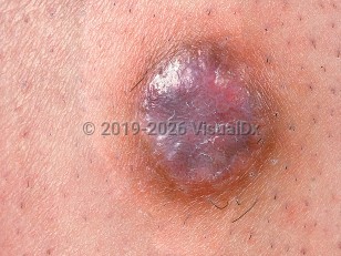
Clinical image of Dermatofibroma - imageId=580299. Click to open in gallery.  caption: 'A close-up of a violaceous papule with a light brown rim.'
A close-up of a violaceous papule with a light brown rim.
Copyright © 2024 VisualDx®. All rights reserved.