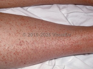
The 3 diagnostic criteria are:
- Peripheral hypereosinophilia (HE) (> 1.5 x 10⁹/L) on 2 occasions separated in time by 4 weeks with or without marked tissue eosinophilia
- HE-related organ damage or dysfunction:
- Greater than 20% eosinophils as compared to all nucleated cells on bone marrow section
- Extensive tissue infiltration by eosinophils as determined by pathologist
- Significant deposition of eosinophil granule proteins in tissue in absence of major tissue infiltration with eosinophils
- Absence of another explanation for the organ damage
- Primary HES – Monoclonal HE due to an underlying stem cell, myeloid, or eosinophilic neoplasm.
- Secondary HES – Polyclonal HE due to an overproduction of eosinophilopoietic cytokines. Potential causes include allergic disorders, malignancy, parasitic infection, and drug reactions.
- Idiopathic HES – Underlying cause of the HE is unknown.
Variants:
- Lymphoproliferative HES is associated with abnormal clones of T-cells producing Th-2 cytokines, specifically interleukin-5, which results in increased eosinophil production. These patients are at increased risk of developing lymphoma.
- Myeloproliferative HES is associated with the FIP1L1-PDGFRA fusion gene (encoding an activated tyrosine kinase), as well other chromosomal aberrations. This genetic mutation is found in various forms of myeloproliferative HES including eosinophilic leukemia.
- Familial HES is associated with autosomal dominant inheritance and linkage to chromosome 5q31-33. Although most affected individuals are asymptomatic, fatal progression of the disease has occurred.
- Other eosinophilic disorders: eosinophilic vasculitis; eosinophilic gastritis, eosinophilic cellulitis; episodic angioedema with eosinophilia (also known as Gleich syndrome); nodules, eosinophilia, rheumatism, dermatitis, and swelling (NERDS) syndrome; etc.
- Myeloproliferative variant (3 types):
- PDGFRA-associated HES (FIP1L1-PDGFRA: interstitial deletion of chromosome 4q12).
- Unknown etiology (FIP1L1-PDGFRA negative) associated HES.
- Chronic eosinophilic leukemia (CEL) characterized by cytogenetic abnormalities and/or peripheral blasts.
- Findings associated with the myeloproliferative variant are hepatosplenomegaly, anemia, thrombopenia, bone marrow dysplasia or fibrosis, and elevated cobalamin levels.
- Lymphocytic variant: Associated with clonal proliferation of T-cells, and primarily characterized by skin disease. This follows a more benign course.
- Idiopathic / undefined variant not associated with chromosomal or clonal abnormalities.
- Overlap variant (organ-restricted eosinophilic disorders).
- Associated variant (eosinophilia in association with a defined diagnosis, for example, Churg-Strauss syndrome).
- Familial variant (familial history of documented persistent eosinophilia of unknown cause).
Worldwide frequency of HES is rare in children and the elderly. FIP1L1-PDGFRA-associated myeloproliferative HES is more common in males in adults, but there is only a slight male predominance in pediatric HES. Age of onset usually ranges from 20-50 years with a mean of 33 years.
Morbidity and mortality from HES is variable. This condition may be quiescent or rapidly fatal from heart failure secondary to progressive restrictive cardiomyopathy. Poorer prognoses are associated with malignant transformation to eosinophilic leukemia, higher eosinophil counts, and cardiac or neurologic involvement.
Children with HES often can present with higher eosinophil counts than adults. However, the clinical presentation, differential diagnosis, treatment, and prognosis are similar to HES in adults.


