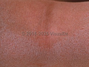
X-linked ichthyosis in Infant/Neonate
Alerts and Notices
Important News & Links
Synopsis

X-linked ichthyosis is an X-linked recessive disorder of abnormal cornification caused by a deficiency of steroid sulfatase, an enzyme that is needed for normal desquamation of the stratum corneum. It is classically characterized by dirty-appearing scale on the sides of the neck ("dirty neck") and the periumbilical area. The remainder of the trunk and extremities are also involved, with classic sparing of the face, palms, and soles, as well as the popliteal and antecubital fossae.
This condition is exclusively seen in males, appears before 3 months of age, and persists for life. During the first few weeks of life there is desquamation of large, loosely adherent, translucent scales followed by the development of tightly adherent, dark brown scales. The disease phenotype ranges from absent to marked and diffuse scaling. It is worse in low-humidity climates.
The disease may be diagnosed prenatally due to a low estriol on a maternal triple screen and confirmed by fluorescence in situ hybridization (FISH). Women with affected fetuses may experience lack of spontaneous labor and prolonged labor, possibly requiring cesarean delivery or vacuum-assisted delivery due to a lack of steroid sulfatase in the placenta, leading to decreased levels of estrogen and insufficient dilation of the cervix.
Codes
ICD10CM:
Q80.1 – X-linked ichthyosis
SNOMEDCT:
72523005 – X-linked ichthyosis with steryl-sulfatase deficiency
Q80.1 – X-linked ichthyosis
SNOMEDCT:
72523005 – X-linked ichthyosis with steryl-sulfatase deficiency
Look For
Subscription Required
Diagnostic Pearls
Subscription Required
Differential Diagnosis & Pitfalls

To perform a comparison, select diagnoses from the classic differential
Subscription Required
Best Tests
Subscription Required
Management Pearls
Subscription Required
Therapy
Subscription Required
References
Subscription Required
Last Reviewed:09/12/2019
Last Updated:01/18/2022
Last Updated:01/18/2022
 Patient Information for X-linked ichthyosis in Infant/Neonate
Premium Feature
VisualDx Patient Handouts
Available in the Elite package
- Improve treatment compliance
- Reduce after-hours questions
- Increase patient engagement and satisfaction
- Written in clear, easy-to-understand language. No confusing jargon.
- Available in English and Spanish
- Print out or email directly to your patient
Upgrade Today

X-linked ichthyosis in Infant/Neonate

