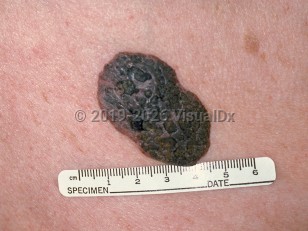
Seborrheic keratoses (SKs) are extremely common, benign neoplasms of the epidermis that typically appear on the chest and back. There can be few or hundreds of these raised, "stuck-on"-appearing papules and plaques with well-defined borders. The etiology is unknown, although there is a familial trait for the development of multiple SKs with an autosomal dominant mode of inheritance.
SKs tend to increase in incidence and number with increasing age. SKs are usually asymptomatic, but when irritated or traumatized, they may become pruritic or painful with associated redness or bleeding.
An SK may start out as a flat, wrinkled thin plaque with a "postage stamp" appearance (flat SK).
A lichenoid keratosis is an inflamed SK that presents as a pink, shiny papule or plaque with an appearance that may resemble that of nodular or cystic basal cell carcinoma (BCC) or melanoma.
Dermatosis papulosa nigra (DPN) is a term given to the small papular SKs (most often seen as dark brown, 1- to 3-mm papules) on the face of individuals with darker skin colors. It is more common in women and can present earlier in life than regular SKs.
Stucco keratoses are smaller, often lighter in color, flat-topped, and scaly, and are common on the lower distal legs of older adult individuals.
Melanoacanthoma is an SK subtype characterized by more hyperpigmentation and less hyperkeratosis and often requires a biopsy for definitive diagnosis given the degree of pigmentation.
Relatively rapid onset (within weeks to months) of numerous SKs may be a cutaneous sign of internal malignancy. Multiple eruptive SKs in association with a visceral cancer is referred to as the sign of Leser-Trélat. The most common associated malignancy is adenocarcinoma of the gastrointestinal tract; other associated malignancies include lung cancer, esophageal carcinoma, mycosis fungoides, and Sézary syndrome.
Seborrheic keratosis
See also in: External and Internal Eye,Anogenital,Hair and ScalpAlerts and Notices
Important News & Links
Synopsis

Codes
ICD10CM:
L82.1 – Other seborrheic keratosis
SNOMEDCT:
25499005 – Seborrheic keratosis
L82.1 – Other seborrheic keratosis
SNOMEDCT:
25499005 – Seborrheic keratosis
Look For
Subscription Required
Diagnostic Pearls
Subscription Required
Differential Diagnosis & Pitfalls

To perform a comparison, select diagnoses from the classic differential
Subscription Required
Best Tests
Subscription Required
Management Pearls
Subscription Required
Therapy
Subscription Required
Drug Reaction Data
Subscription Required
References
Subscription Required
Last Reviewed:07/28/2025
Last Updated:08/04/2025
Last Updated:08/04/2025
 Patient Information for Seborrheic keratosis
Premium Feature
VisualDx Patient Handouts
Available in the Elite package
- Improve treatment compliance
- Reduce after-hours questions
- Increase patient engagement and satisfaction
- Written in clear, easy-to-understand language. No confusing jargon.
- Available in English and Spanish
- Print out or email directly to your patient
Upgrade Today

Seborrheic keratosis
See also in: External and Internal Eye,Anogenital,Hair and Scalp