Hemophagocytic lymphohistiocytosis (HLH) requires evaluation and management by a team of specialist physicians with experience in the disease, including but not limited to hematologist / oncologists and infectious disease specialists. While definitive management should be targeted at the underlying etiology, steroids are commonly used to initially stabilize those with severe disease. Without treatment, HLH can be rapidly fatal.
Diagnosis Overview:
Hemophagocytic lymphohistiocytosis (HLH), also known as histiocytic medullary reticulosis, macrophage activation syndrome, and familial hemophagocytic lymphohistiocytosis (for inherited forms), is a relatively uncommon hemophagocytic syndrome. HLH affects all age groups, but the majority of cases are in infants (often younger than 1 year) and young children. The secondary form is more common in older children and adults.
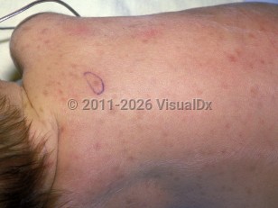
HLH tends to affect the bone marrow, spleen, lymph nodes, liver, central nervous system, and skin. Up to 65% of patients have nonspecific cutaneous involvement ranging from a transient generalized morbilliform rash to petechiae, purpuric macules, or papules to erythroderma.
Other common clinical signs include hepatomegaly, lymphadenopathy, nonspecific cutaneous eruptions, and neurological abnormalities.
It is classified into primary (familial) and secondary (acquired, sporadic) forms:
- Familial hemophagocytic lymphohistiocytosis (FHL) is inherited in an autosomal recessive pattern; approximately 24% of 122 cases reviewed by the FHL Study Group of the Histiocyte Society had a history of parental consanguinity. Estimated incidence of FHL is 1.2 per million in children under the age of 15; 70%-80% of FHL cases present before age 1 year. Up to 80% of FHL cases are caused by mutations in 1 of 3 genes (encoded protein in parentheses): PRF-1 (perforin, most common), UNC13D (Munc13-14), and STX-11 (syntaxin-11). Both perforin and Munc13-14 proteins are involved in host response to infection, while the exact role of syntaxin-11 has yet to be determined.
- Secondary HLH may occur sporadically or in the context of FHL. It is thought to be related to immune stimulation by infection (eg, Epstein-Barr virus [EBV], human herpes simplex virus [HSV], coxsackievirus B, echovirus, other human herpesviruses, tuberculosis, and other viral, fungal, and parasitic infections), malignancy (eg, acute lymphoblastic leukemia, acute myelogenous leukemia, and lymphomas), collagen vascular disease (eg, juvenile rheumatoid arthritis), and immunodeficiency (eg, HIV). EBV-associated HLH may present similarly to a T-cell lymphoma. Human HSV-, coxsackievirus B-, and echovirus-associated HLH should be a consideration in neonates. HLH has also been reported in association with Chediak-Higashi, Griscelli, and rare X-linked lymphoproliferative syndromes.
While infections (including but not limited to viral infection like EBV and cytomegalovirus [CMV] or bacterial infections) tend to be the more common trigger in children and adolescents, malignancy (lymphoma, leukemia) tends to be the more common cause in adults. Autoimmune disease (systemic lupus erythematosus, adult-onset Still disease) is also often a cause.
The pathophysiology behind both primary and secondary HLH is T-cell immune dysregulation. While the exact mechanism is unknown, the dysregulatory process results in an overproduction of inflammatory cytokines such as tumor necrosis factor-alpha (TNF-alpha), interferon gamma (IFN-gamma), and various interleukins (IL). These molecules activate T-cells, macrophages, and histiocytes. This disrupts the mechanisms that regulate apoptosis. Natural killer (NK) cells, particularly in FHL, are absent or reduced. Under normal circumstances, NK cells (which have granules containing perforin and granzymes that disrupt target cell membrane integrity) participate in natural cell death.
HLH-2004 diagnostic criteria require 5 of 8 criteria for diagnosis. A 2024 revision addresses primary (familial) HLH; it excludes NK cell activity and requires 5 of the remaining 7 criteria.
- Fever
- Splenomegaly
- Cytopenias (greater than or equal to 2 of 3 lineages below)
- Hemoglobin: less than 90 g/L (in neonates, less than 100 g/L)
- Platelets: less than 100 x 109/L
- Neutrophils: less than 109/L (in absence of hypocellular marrow)
- Hypertriglyceridemia (fasting levels greater than or equal to 3 mmol/L) and/or hypofibrinogenemia (fibrinogen level of less than or equal to 1.5 g/L)
- Tissue sample (from bone marrow, liver, or lymph nodes) demonstrating macrophage phagocytosis of erythrocytes, platelets, leukocytes, and precursor cells
- Low / absent NK activity
- Hyperferritinemia (greater than or equal to 500 μg/L)
- High levels of soluble IL-2 receptor (soluble CD25 greater than or equal to 2400 U/mL)
Immunocompromised Patient Considerations: HLH can be triggered by infections that lead to immunodeficiency states such as HIV infection or by noninfectious conditions that lead to immunodeficiency states like hematologic malignancies.