Sarcoidosis - Hair and Scalp
See also in: Overview,External and Internal Eye,Oral Mucosal LesionAlerts and Notices
Important News & Links
Synopsis

Sarcoidosis commonly presents with abnormalities identified incidentally on chest radiography. Although the disease can affect different organs, systemic symptoms such as fever, night sweats, and weight loss are common. Sarcoidosis can affect the lungs, with symptoms such as chest pain, shortness of breath, cough, and most commonly, pronounced fatigue. It can also affect the peripheral lymph nodes, heart, kidneys, gastrointestinal tract, central nervous system (CNS), liver, spleen, bone, muscle, and endocrine glands. Venous thromboembolism and pulmonary hypertension are potential complications of sarcoidosis. Approximately 90% of patients will have lung involvement. Pulmonary fibrosis and bronchiolectasis result in "honeycombing" of the lung and represent end-stage lung disease due to chronic granulomatous inflammation. Hilar lymphadenopathy is asymptomatic and affects 90% of patients. Approximately 10% of patients have hypercalcemia.
Neurosarcoidosis commonly manifests as cranial-nerve deficits. Any part of the neurologic system can be impacted by sarcoidosis; a vast range of neurologic signs and symptoms may be present. Delays in diagnosis and treatment can lead to permanent disability. The diagnostic workup for neurosarcoidosis should include an MRI of the head, cerebrospinal fluid analysis, and detection of sarcoidosis outside the nervous system.
Approximately 25% of patients will have cutaneous involvement, and some patients may have skin-limited disease. Asymptomatic red-brown dermal papules and/or plaques that favor the face, neck, upper extremities, and upper trunk are the most common specific cutaneous findings. Less common manifestations include sarcoid lesions with epidermal change such as scale (ichthyosiform sarcoid), hypopigmentation, subcutaneous nodules, and ulceration. Sarcoidosis has a predilection for scars and may be seen within tattoos.
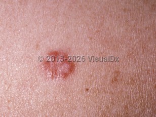
Sarcoidosis of the scalp is a rarely reported cutaneous finding. Both nonscarring and scarring alopecia may occur in association. Scarring plaques are usually localized, and nonscarring alopecia may be localized or generalized. Underlying plaques may be red-brown, violaceous, and/or hypopigmented. They may be raised or depressed and atrophic. Scaling can be seen, and scalp involvement can resemble seborrheic dermatitis. An annular morphology has been reported. Most reported cases have been seen in African American women. Scalp sarcoid has been correlated with the presence of systemic involvement. Scalp sarcoid can be recalcitrant to treatment.
The pathogenesis of sarcoidosis is poorly understood. However, it is characterized by noncaseating epithelioid granulomas made up mostly of CD4+ helper T-cells, a predominantly Th1 type immune response, and elevated levels of interferon (IFN)-gamma and interleukin (IL)-2.
Codes
D86.3 – Sarcoidosis of skin
SNOMEDCT:
31541009 – Sarcoidosis
Look For
Subscription Required
Diagnostic Pearls
Subscription Required
Differential Diagnosis & Pitfalls

Subscription Required
Best Tests
Subscription Required
Management Pearls
Subscription Required
Therapy
Subscription Required
Drug Reaction Data
Subscription Required
References
Subscription Required
Last Updated:11/10/2022
 Patient Information for Sarcoidosis - Hair and Scalp
- Improve treatment compliance
- Reduce after-hours questions
- Increase patient engagement and satisfaction
- Written in clear, easy-to-understand language. No confusing jargon.
- Available in English and Spanish
- Print out or email directly to your patient


