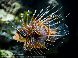
Lionfish spine puncture
Alerts and Notices
Important News & Links
Synopsis

Lionfish include several species of venomous fish belonging to the large Scorpaenidae family. Members of the Scorpaenidae family can be divided into 3 groups based on their venom organs and toxicity: lionfish (Pterois), scorpionfish (Scorpaena), and stonefish (Synanceja). Scorpaenidae stings are progressively more severe from lionfish to scorpionfish to stonefish. Lionfish typically have long, relatively slender spines with the smallest venom glands, and they produce the weakest venom. Scorpionfish have shorter, but sturdier spines and larger venom glands, and, thus, have the potential to deliver a more potent sting. Stonefish have the shortest and strongest spines, the largest venom glands, and can deliver a much larger dose of far more powerful venom to a victim. Each spine is covered with a loose integumentary sheath. During envenomation, the sheath is pushed down the spine, causing compression of the venom glands located at the base of the spines. Venom then travels through grooves in the spines and into the wound.
Lionfish are notable for their extremely long and separated spines, and they are generally striped in appearance. There are typically 12-13 dorsal spines, 2 pelvic spines, and 3 anal spines, all venomous. They also have nonvenomous ornate pectoral spines. They inhabit tropical waters and, less commonly, temperate waters. In the United States, they are common aquarium pets and inhabit the Atlantic Ocean from North Carolina to South Florida.
Lionfish venom is heat labile and affects neuromuscular transmission.
In the United States, lionfish envenomation typically occurs to the upper extremities of aquarist by captive lionfish. Severe, sharp local pain is the predominant symptom, and the pain may radiate throughout the affected limb. If untreated, the pain worsens over the next 1-2 hours and typically persists for 6-12 hours, though an ache can last for weeks. Subsequent erythema, edema, and warmth may involve the affected limb. The wound area is initially ischemic and then cyanotic. Vesicles may form followed by tissue sloughing with surrounding cellulitis. Necrotic ulceration is rare. The intensity of the sting depends on the size of the fish and then the number of spines penetrating the skin, as well as other factors, such as body weight and health of the victim.
Lionfish envenomation wounds are classified into 3 grades: Grade I (erythema), Grade II (vesicle formation), and Grade III (tissue necrosis). Most envenomations are Grade I.
Systemic symptoms occur in approximately 13% of lionfish envenomations (according to two studies). The most common symptoms include nausea, diaphoresis, chills, muscle weakness, shortness of breath, hypotension, abdominal pain, and syncope.
Late complications include paresthesias, secondary infection, ulceration, granuloma formation, or fibrous soft-tissue defects. Hypersensitivity to lionfish venom may cause an allergic reaction on subsequent envenomation.
Codes
ICD10CM:
T63.591A – Toxic effect of contact with other venomous fish, accidental (unintentional), initial encounter
SNOMEDCT:
241824004 – Poisoning from lion fish spine
T63.591A – Toxic effect of contact with other venomous fish, accidental (unintentional), initial encounter
SNOMEDCT:
241824004 – Poisoning from lion fish spine
Look For
Subscription Required
Diagnostic Pearls
Subscription Required
Differential Diagnosis & Pitfalls

To perform a comparison, select diagnoses from the classic differential
Subscription Required
Best Tests
Subscription Required
Management Pearls
Subscription Required
Therapy
Subscription Required
References
Subscription Required
Last Updated:10/18/2017

