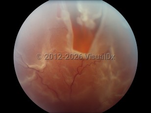
Retinal tear - External and Internal Eye
Alerts and Notices
Important News & Links
Synopsis

Retinal tears are associated with myopia (nearsightedness), pseudophakia (after cataract surgery), lattice degeneration, and trauma. Liquefaction of the vitreous and posterior vitreous detachments lead to vitreous traction and may lead to a break in the retina. Blunt trauma to the eye may rapidly compress the eye in the anterior-posterior plane and expand the eye in the equatorial plane, causing severe traction and possible retinal breaks.
When the vitreous fluid leaks through the tear, fluid can accumulate beneath the retina and separate the retina from the underlying retinal pigment epithelium and the choriocapillaris (blood supply). This separation of layers is called a retinal detachment. Most retinal breaks do not progress to retinal detachment.
Symptoms of floaters and flashing lights may accompany a retinal tear, though most breaks remain asymptomatic. A retinal tear (especially symptomatic ones) may progress to a retinal detachment within hours, leading to progressive visual field loss.
Codes
H33.039 – Retinal detachment with giant retinal tear, unspecified eye
H33.319 – Horseshoe tear of retina without detachment, unspecified eye
SNOMEDCT:
95690009 – Retinal tear
Look For
Subscription Required
Diagnostic Pearls
Subscription Required
Differential Diagnosis & Pitfalls

Subscription Required
Best Tests
Subscription Required
Management Pearls
Subscription Required
Therapy
Subscription Required
Drug Reaction Data
Subscription Required
References
Subscription Required

