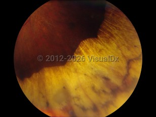
Retinopathy of prematurity - External and Internal Eye
Alerts and Notices
Important News & Links
Synopsis

Retinopathy of prematurity (ROP) is a proliferative retinopathy of premature and low-birth-weight infants. This disease was termed retrolental fibroplasia in the past.
Incomplete vascularization of the retina triggers development of ROP. The extent of the vascularity of the retina mainly depends on how early the baby is born. Starting at 16 weeks' gestation, blood vessels normally spread from the optic disc to the peripheral retina. By term, mature blood vessels extend fully to the peripheral retina. The avascular peripheral retina in ROP produces vascular growth factors that can, in turn, induce pathological neovascularization and may lead to retinal detachment.
The major risk factors for ROP are prematurity and low birth weight. White and Hispanic children tend to develop a more aggressive ROP that is more likely to cause blindness. Supplemental oxygen therapy plays a role for premature infants. When withdrawn from the oxygen, the avascular retina will produce vascular endothelial growth factor (VEGF), inducing the blood vessel growth.
Because there are no symptoms of ROP, high-risk babies need to be screened with dilated fundus examinations by experienced ophthalmologists. ROP can progress rapidly over days to weeks, but most cases of ROP regress spontaneously by a process of involution.
While screening and early intervention of ROP have improved over the decades, there has also been an increased survival of lower-birth-weight infants. ROP is now the leading cause of childhood blindness in industrialized countries and is the fifth leading cause of total childhood blindness worldwide.
Codes
ICD10CM:
H35.109 – Retinopathy of prematurity, unspecified, unspecified eye
SNOMEDCT:
415297005 – Retinopathy of prematurity
H35.109 – Retinopathy of prematurity, unspecified, unspecified eye
SNOMEDCT:
415297005 – Retinopathy of prematurity
Look For
Subscription Required
Diagnostic Pearls
Subscription Required
Differential Diagnosis & Pitfalls

To perform a comparison, select diagnoses from the classic differential
Subscription Required
Best Tests
Subscription Required
Management Pearls
Subscription Required
Therapy
Subscription Required
References
Subscription Required
Last Updated:07/11/2013

