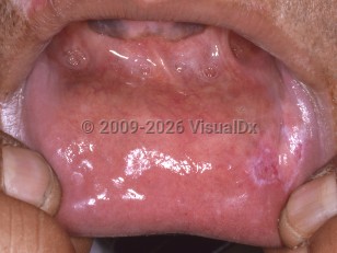
Oral lupus erythematosus - Oral Mucosal Lesion
Alerts and Notices
Important News & Links
Synopsis

Lupus erythematosus is a chronic inflammatory autoimmune disease that can manifest cutaneously, orally, and systemically. Risk factors for developing cutaneous lesions include sex (3:1 female-to-male ratio, especially during childbearing years) and race/ethnicity, with Black patients demonstrating a higher incidence compared with White patients. Cutaneous lupus erythematosus may be acute, subacute, or chronic (also known as discoid). Sex and race/ethnicity are also risk factors for developing systemic lupus erythematosus (SLE), with a 6:1 female-to-male ratio, and with Black women demonstrating a fourfold higher incidence compared with White women. Individuals of childbearing potential are most commonly affected. Lupus erythematosus can also be drug induced.
Oral mucosal lesions can occur in both cutaneous and systemic forms of lupus erythematosus. The lips, gingiva, tongue, and palatal and buccal mucosa are the most commonly involved sites.
For a more in-depth discussion of the subtypes of cutaneous lupus erythematosus and of SLE, see Discoid lupus erythematosus, Subacute cutaneous lupus erythematosus, and Systemic lupus erythematosus. See also Drug-induced lupus erythematosus.
Codes
ICD10CM:
L93.2 – Other local lupus erythematosus
SNOMEDCT:
403495008 – Discoid lupus erythematosus of oral mucosa
L93.2 – Other local lupus erythematosus
SNOMEDCT:
403495008 – Discoid lupus erythematosus of oral mucosa
Look For
Subscription Required
Diagnostic Pearls
Subscription Required
Differential Diagnosis & Pitfalls

To perform a comparison, select diagnoses from the classic differential
Subscription Required
Best Tests
Subscription Required
Management Pearls
Subscription Required
Therapy
Subscription Required
References
Subscription Required
Last Updated:08/16/2021
Oral lupus erythematosus - Oral Mucosal Lesion

