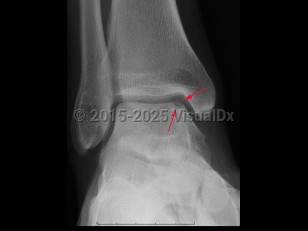
Osteochondral defect
Alerts and Notices
Important News & Links
Synopsis
An osteochondral defect is a rare condition of osteonecrosis of the subchondral bone, characterized by a loose bone fragment and articular cartridge which is partially or completely detached from underlying bone. Joints most commonly affected are knee, elbow, and ankle. Symptoms include localized pain following athletic injury or overuse, more often in boys than girls. If the fragment becomes lodged in the joint, the patient may experience increased pain, tenderness, locking, or crepitus of the joint. Treatment depends on growth stage of affected patient. Juveniles with open growth plates may be treated conservatively and nonsurgically. Adults who are skeletally mature may undergo arthroscopic repair.
Related topics: osteochondral defect of talus, osteochondritis dissecans of elbow, osteochondritis dissecans of knee
Related topics: osteochondral defect of talus, osteochondritis dissecans of elbow, osteochondritis dissecans of knee
Codes
ICD10CM:
M93.20 – Osteochondritis dissecans of unspecified site
SNOMEDCT:
82562007 – Osteochondritis dissecans
M93.20 – Osteochondritis dissecans of unspecified site
SNOMEDCT:
82562007 – Osteochondritis dissecans
Best Tests
Subscription Required
References
Subscription Required
Last Updated:09/11/2023
Osteochondral defect

