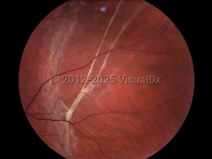
Retinal detachment - External and Internal Eye
Alerts and Notices
Important News & Links
Synopsis

Most retinal detachments are rhegmatogenous, caused by a break in the retina. Age, surgical and nonsurgical trauma, high myopia, and peripheral retinal degeneration predispose to these breaks. A posterior vitreous detachment (PVD) is usually a benign condition associated with age and liquefaction of the vitreous, but its tractional force may lead to retinal breaks. While most retinal breaks do not progress to a retinal detachment, the prophylactic treatment available and the potential for vision loss signifies the importance of detecting retinal breaks.
Retinal detachments can also be non-rhegmatogenous, caused either by leakage from beneath the retina (exudative) or by traction from scar tissue on the retina (tractional). Proliferative diabetic retinopathy, proliferative vitreoretinopathy, retinopathy of prematurity, sickle cell retinopathy, and retinal vein occlusion can be associated with vitreous membranes that pull the neurosensory retina from the RPE.
Exudative detachments result when damaged RPE or retinal blood vessels allow fluid to pass into the subretinal space. It can be associated with ocular inflammation (uveitis, scleritis), neoplasms (choroidal melanoma, metastatic disease), vascular disease (Coats' disease, malignant hypertension, eclampsia), maculopathies (central serous chorioretinopathy), and congenital disorders (optic disc pit, nanophthalmos). In exudative retinal detachments, subretinal fluid shifts with head position, and visual disturbances may be positional.
Patients who see photopsias (flashes of light) or floaters are at an increased risk of developing retinal breaks and detachments. There should be a high suspicion of progression to retinal detachment when bright flashes are accompanied by a shower of black dots or a curtain or black shadow blocking the vision.
The overall annual incidence of retinal detachments is approximately 1 in 10,000. Although most detachments can be repaired with anatomic success, only those treated early avoid permanent visual impairment. Retinal detachments can progress to involve the macula within hours, often leading to visual loss even when repaired properly.
Codes
H33.009 – Unspecified retinal detachment with retinal break, unspecified eye
SNOMEDCT:
42059000 – Retinal detachment
Look For
Subscription Required
Diagnostic Pearls
Subscription Required
Differential Diagnosis & Pitfalls

Subscription Required
Best Tests
Subscription Required
Management Pearls
Subscription Required
Therapy
Subscription Required
Drug Reaction Data
Subscription Required
References
Subscription Required

