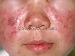
The etiology of SLE is poorly understood, but there is a strong association with autoantibodies and SLE. For example, even though the autoantibodies are not organ specific, only certain organs in a given patient demonstrate end-organ damage. It is hypothesized that a complex interplay between genetic proclivity and environmental influences leads to a perpetuated autoimmune response.
Autoantibodies play significant roles in the diagnosis, management, and prognosis of SLE. They are as follows:
- Anti-dsDNA – Highly specific for SLE. Rising levels correlate with increased SLE activity and an increased risk for SLE nephritis. Seen in approximately 55%-65% of SLE patients.
- Anti-Smith (anti-Sm) – Highly specific for SLE. Seen in approximately 25%-30% of SLE patients. Considerable diagnostic value, but levels do not correlate with disease activity.
- Anti-RNP – Highly specific for SLE. Seen in approximately 5% of SLE patients.
- Antinuclear antibody (ANA) – Highly sensitive for SLE. Seen in approximately 99% of SLE patients. In other words, it is very rare for an individual with SLE to have a negative ANA. Considerable screening value, but levels do not correlate with disease activity.
- Anti-histones – Highly specific for drug-induced SLE.
The organ systems most commonly affected in SLE are the joints, skin, renal system, pulmonary system, central nervous system (including ischemic stroke), cardiovascular system, and hematologic system. Patients may be anemic. About half of SLE patients will have significant renal involvement that can take the form of several types of glomerulonephritis. A classification system is available from the International Society of Nephrology / Renal Pathology Society, in which most cases are class II through V. Classes III and IV have a poor prognosis. Class V is also known as membranous lupus nephritis. A very significant percentage of patients with membranous lupus nephritis and nephrotic proteinuria will progress to end-stage kidney disease. Renal biopsy is useful to define the type and extent of involvement and to guide therapy. Other complications include thromboembolic disease, particularly in the setting of antiphospholipid antibodies and vasculitis, and gastrointestinal (GI), pulmonary, and cardiac involvement. Choreiform movements may occur.
Skin and joint findings:
- Arthritis is classically migratory, polyarticular, and symmetrical and a common early finding.
- Skin and mucous membrane abnormalities are also common with the classic "butterfly" malar rash (involving the cheeks and nose) developing after sun exposure – This form of cutaneous lupus is sometimes referred to as acute cutaneous lupus erythematosus (ACLE). There is a range of other skin findings that can occur with SLE. For more details, see subacute cutaneous lupus erythematosus, discoid lupus erythematosus, tumid lupus erythematosus.
- Bullous systemic lupus erythematosus is a rare autoimmune blistering disease in patients with a known diagnosis of SLE.
- Lupus profundus is a rare panniculitis seen in patients with SLE.
- Nonbullous neutrophilic lupus erythematosus is an extremely rare eruption of widespread urticarial papules and plaques that has been reported in patients with SLE.
SLE is a chronic disease with no known cure. However, there are several disease-modifying medications that are effective in decreasing the burden of disease. The mortality from SLE has decreased in the last several decades. Certain patient characteristics portend a worse prognosis in SLE: male sex, age (both young and old), low socioeconomic status, and being of African descent. Disease phenotypes associated with a poor prognosis include hypertension, renal involvement, antiphospholipid antibody positivity, and antiphospholipid antibody syndrome.
The presence of erythema multiforme-like lesions in a patient with lupus, along with a speckled pattern of antinuclear antibody (ANA), positive anti-Ro/SSA or anti-La/SSB, and positive rheumatoid factor (RF) is known as Rowell syndrome. This syndrome has been described in patients with discoid lupus erythematosus (DLE), subacute cutaneous lupus erythematosus (SCLE), and SLE. Its existence as a distinct entity has been debated in the literature; some authors believe the association is coincidental. Prednisone with or without hydroxychloroquine, dapsone, or immunosuppressive drugs such as cyclosporin have been cited as therapy.
Related topic: neonatal lupus erythematosus


 Patient Information for
