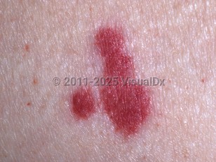
The diagnosis of vasculitis can be challenging as many symptoms are nonspecific and nondiagnostic. The diagnosis is usually made through a combination of a thorough history, physical examination, and laboratory studies, imaging, and biopsy studies. Vasculitis is an uncommon condition in the pediatric population, with an annual incidence of 1.2-5.3 cases per 10 000 children younger than 17 years.
It is important to distinguish a primary systemic vasculitis from one associated with medications, infection, malignancy, or a connective tissue disorder, as the best course of treatment may differ. Drug-induced vasculitis should be quickly recognized and managed by removing the causative medication.
There are several vasculitis classification systems that are based on the size of blood vessels predominately involved (small, medium, or large vessel), histopathology (granulomatous, nongranulomatous, or immuncomplex), clinical manifestations (single organ or multiorgan), and laboratory findings (ANCA positive). While there is no uniform classification used, the most commonly employed pediatric specific classification system is proposed by the European League against Rheumatism (EULAR) and the Pediatric Rheumatology European Society (PReS), which is primarily based on the size of the involved vessels.
There are several different recognized vasculitic disorders in pediatrics.
Predominately large-vessel vasculitis:
Takayasu arteritis – An immune-mediated vasculitis primarily involving the aorta and its branches. Initial manifestations are nonspecific and include fever, weight loss, and joint aches. As the disease progresses, symptoms related to the area of the body to which blood flow is diminished begin (eg, diminished carotid arteries lead to headaches, diminished mental capacity, and confusion).
Predominantly medium-vessel vasculitis:
- Childhood-onset polyarteritis nodosa (PAN) – An immune-mediated vasculitis with initial symptoms of fevers, weight loss, joint aches, myalgias, and skin manifestations. The lower extremities are typically involved, with painful cutaneous and subcutaneous nodules. Lesions can ulcerate, bullae can form, and, rarely, there can be purpura or gangrene. Livedo reticularis is a common associated finding as are nail fold infarctions. Over time, many organ systems are affected.
- Kawasaki disease – A vasculitis of unknown etiology, usually affecting children younger than 5 years. It begins with fevers over 5 days, skin rash, strawberry tongue, swollen hands / feet, red cracked lips, and nonexudative conjunctivitis. If not treated, aneurysms may develop, including of the coronary arteries.
- Cutaneous PAN – A rare immune-mediated vasculitis affecting dermal and subcutaneous vessels without systemic involvement. Manifestations include painful tender nodules, livedo reticularis, and skin ulcers.
- Granulomatosis
- Granulomatosis with polyangiitis (GPA; formerly Wegener granulomatosis) – A rare autoimmune vasculitis involving granuloma formation primarily affecting the respiratory tract and kidneys, although it can involve other organ systems. Manifestations include cough, nasal congestion, epistaxis, sinus pain and pressure, and shortness of breath / increased work of breathing. Renal manifestations can range from hematuria / proteinuria to renal failure. Skin findings include palpable purpura and nodules, followed by necrotic ulcerations, most commonly on the lower extremities.
- Eosinophilic granulomatosis with polyangiitis (EGPA; formerly Churg-Strauss syndrome) – A rare vasculitis involving eosinophilic infiltration of affected tissues. This can involve any organ system with symptoms based on the system involved. Peripheral eosinophilia with asthma (wheezing / cough) and rhinosinusitis are common associations. Cutaneous manifestations include erythema, urticarial plaques, palpable purpura, retiform purpura, ecchymoses, livedo racemosa, necrotic lesions, and tender subcutaneous nodules.
- Nongranulomatosis
- Immunoglobulin A (IgA) vasculitis (formerly Henoch-Schönlein purpura) – An IgA immune-mediated vasculitis involving the small vessels of the skin, kidneys, gastrointestinal (GI) tract, and joints. Common manifestations include petechiae and palpable purpura, primarily of the lower extremities / buttocks, abdominal pain, vomiting, and proteinuria / hematuria.
- Cutaneous leukocytoclastic vasculitis (hypersensitivity vasculitis) – Manifestations are usually limited to the skin and include palpable purpura, bullae, and ulcers, usually involving the lower extremities.
- Acute hemorrhagic edema of infancy is a leukocytoclastic vasculitis affecting children aged 4 months to 2 years. It is characterized by edematous hemorrhagic lesions of the head and distal extremities and is usually noted after an upper respiratory tract infection and/or a course of antibiotics.
- Hypocomplementemia urticarial vasculitis – A rare immune-complex mediated vasculitis that causes recurrent urticarial papules and plaques, which may contain foci of purpura, along with low blood complement levels (C1q). Systemic involvement is common, resulting in asthma-like symptoms, arthritis, and abdominal pain.
- Microscopic polyangiitis – ANCA-positive small vessel vasculitis primarily involving the lungs and kidneys but can involve the GI tract and skin. Manifestations include fever, weight loss, joint aches, palpable purpura, glomerulonephritis, hemoptysis, shortness of breath / increased work of breathing, mononeuritis multiplex, and abdominal pain.
- Behçet disease
- Cogan syndrome
- Vasculitis associated with a connective tissue disease
- Vasculitis associated with malignancy
- Vasculitis associated with a drug
- Vasculitis associated with an infection (hepatitis B, hepatitis C, influenza, HIV, parvoviral infections, Neisseria spp, mycobacterial infections, and Streptococcus spp)
Prognosis is highly variable and is determined by the individual disease, severity of the disease, and the patient's clinical status. Early diagnosis and intervention (if needed) usually improves prognosis.


